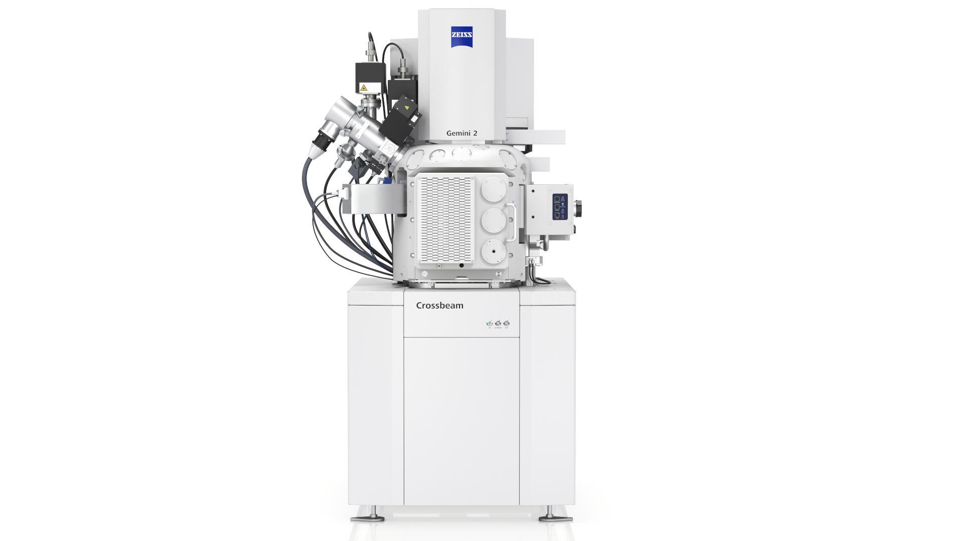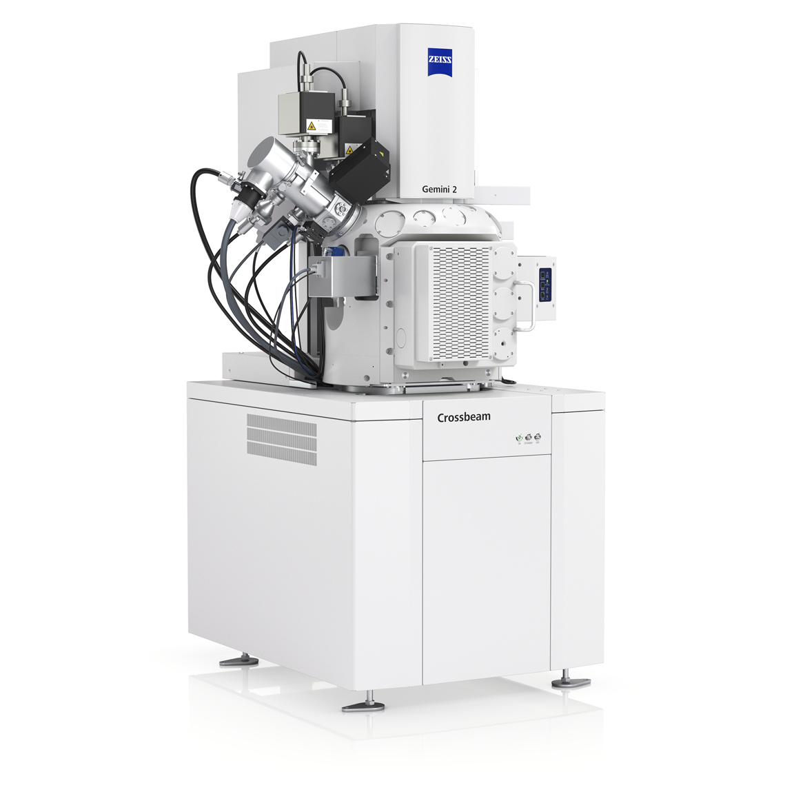

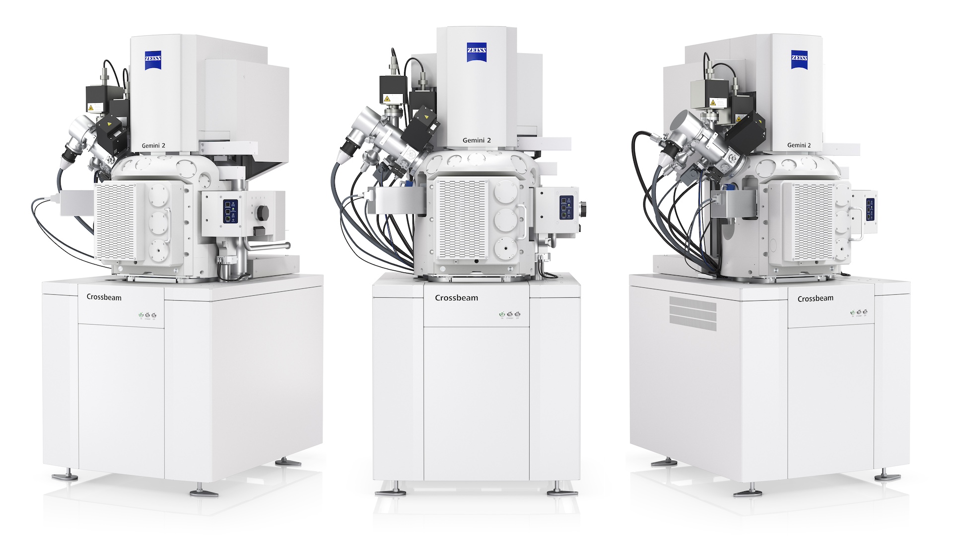
ZEISS Crossbeam
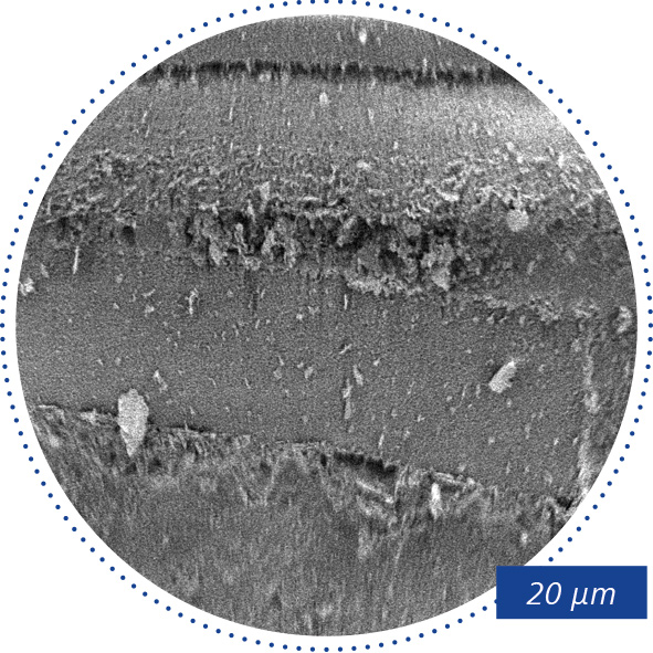
Zaměřen na třetí dimenziKombinace rastrovacího elektronového mikroskopu (SEM) a fokusovaného iontového paprsku (FIB) umožňuje cílené řezání do materiálu v nejmenším měřítku (nanometrový rozsah) a přímo zobrazovat strukturu materiálu pod povrchem. Mezi typické aplikace patří přesná lokalizace a chemická analýza (EDS) lokálních defektů.
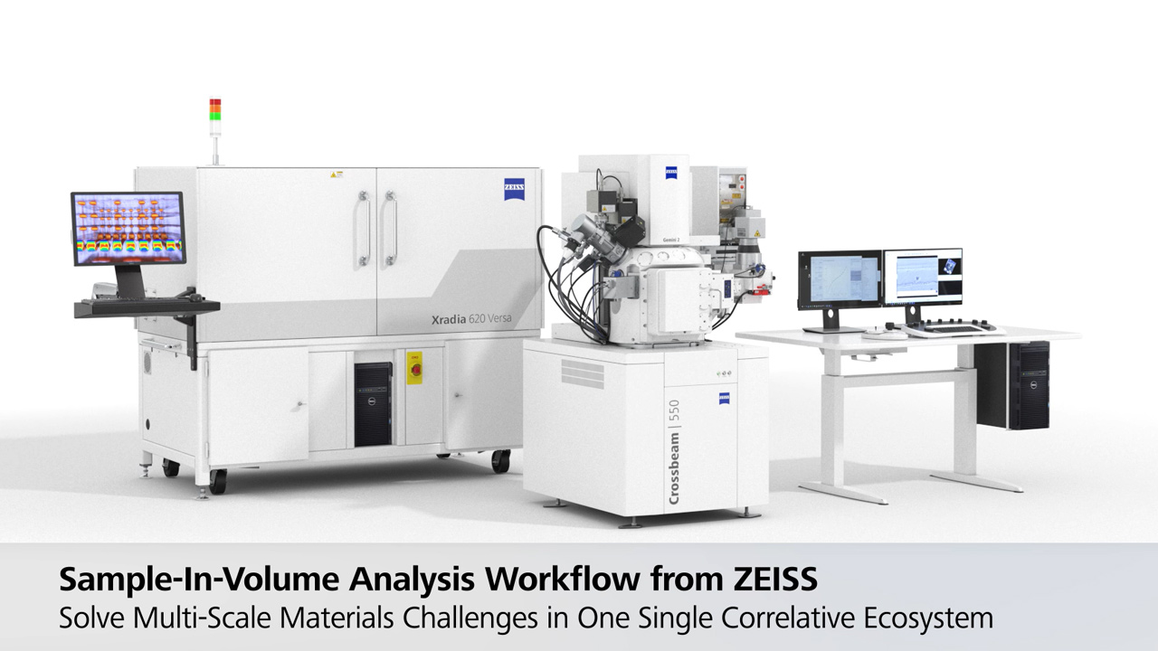
ZEISS Crossbeam pro průmysl
Zažijte novou úroveň testování vašich vzorků.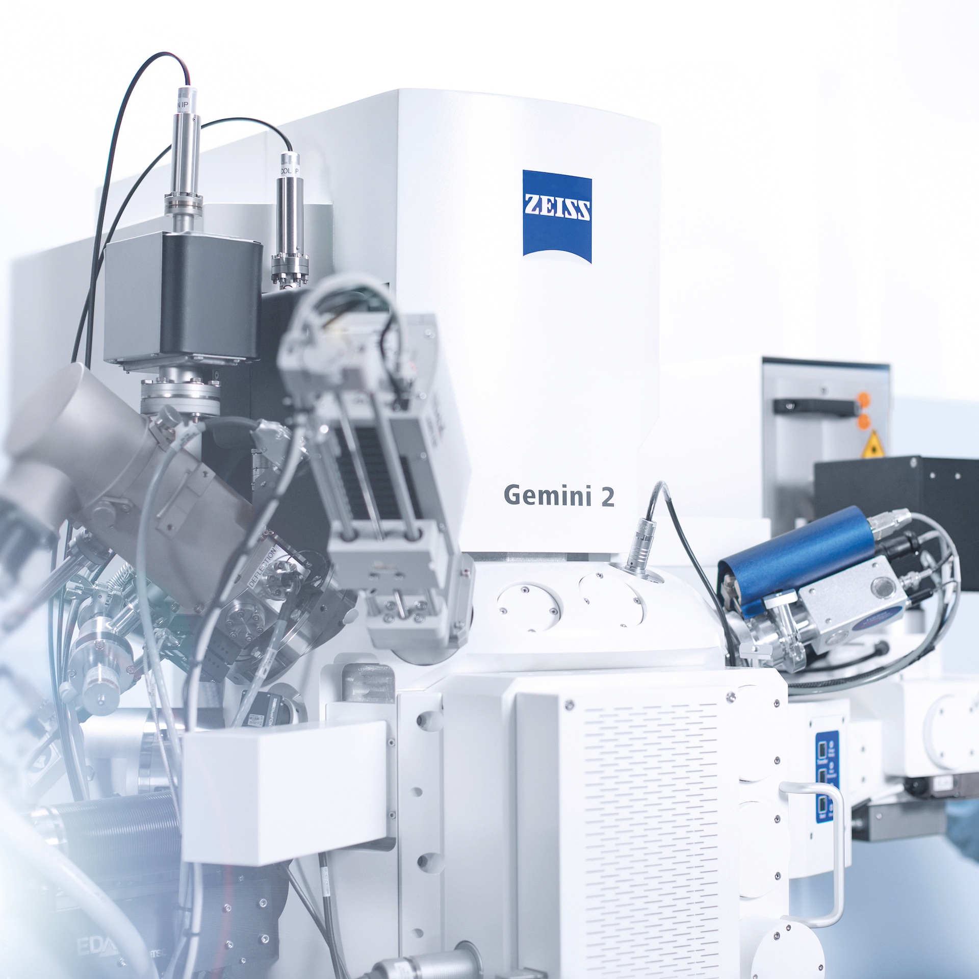

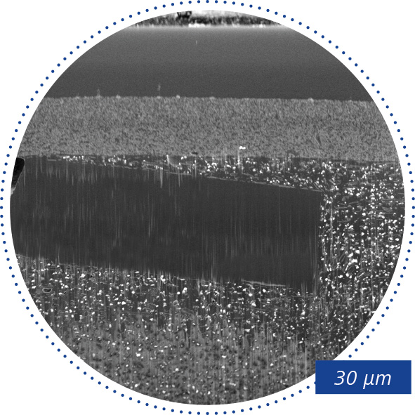
Připravte si tenké lamely pro jejich analýzu v TEM (transmisní elektronová mikroskopie) nebo STEM (rastrovací transmisní elektronová mikroskopie). ZEISS Crossbeam nabízí kompletní řešení pro přípravu TEM lamel i v dávkách.
Nízkonapěťový výkon kolony ion-sculptor FIB podporuje vysoce kvalitní lamely a zabraňuje amorfizaci jemných vzorků. Pro zahájení práce použijte jednoduchý pracovní postup a počkejte na automatické spuštění. Využijte výhod softwaru pro detekci koncových bodů, který poskytuje přesné informace o tloušťce vaší lamely.
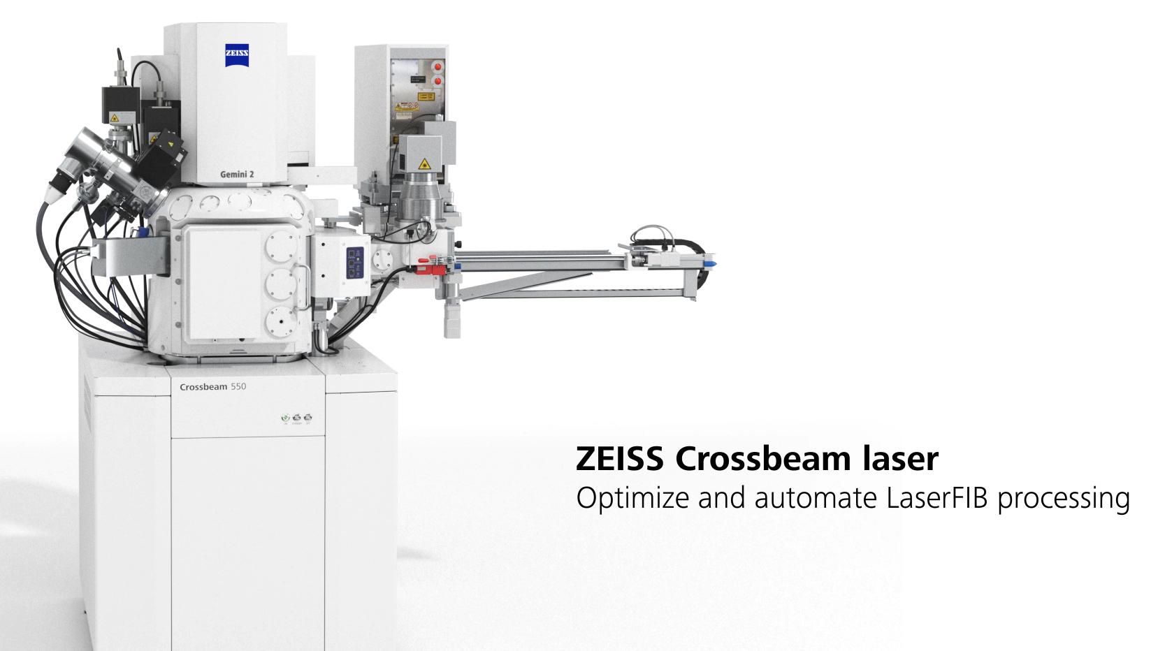
Volitelný femtosekundový laser se používá pro odebrání materiálu a lepší přístup k hlubším strukturám, stejně jako pro přípravu velkých vzorků.

Oblasti použití
- Lokální průřezy, např. v místech defektů (defekty růstu tenkých vrstev, koroze, zachycené částice atd.)
- Příprava lamel pomocí TEM
- Vysokorozlišovací průřezová zkoumání s transmisní spektroskopií (STEM)
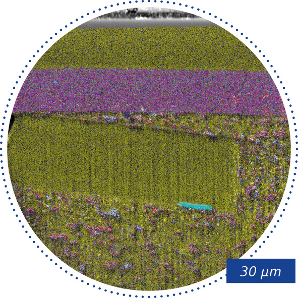
- 3D tomografie mikrostruktury nebo lokálních defektů
- Zpracování struktur cíleným odstraňováním materiálu
Zjistěte více v našich videích o mikroskopu ZEISS Crossbeam
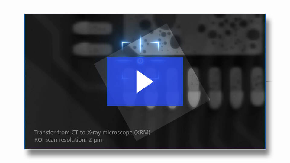


Analýza poruch FIB-SEM na dílech karoserie


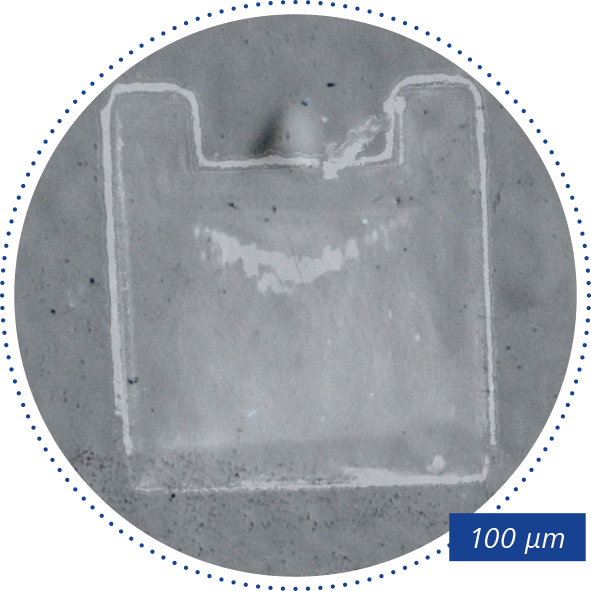

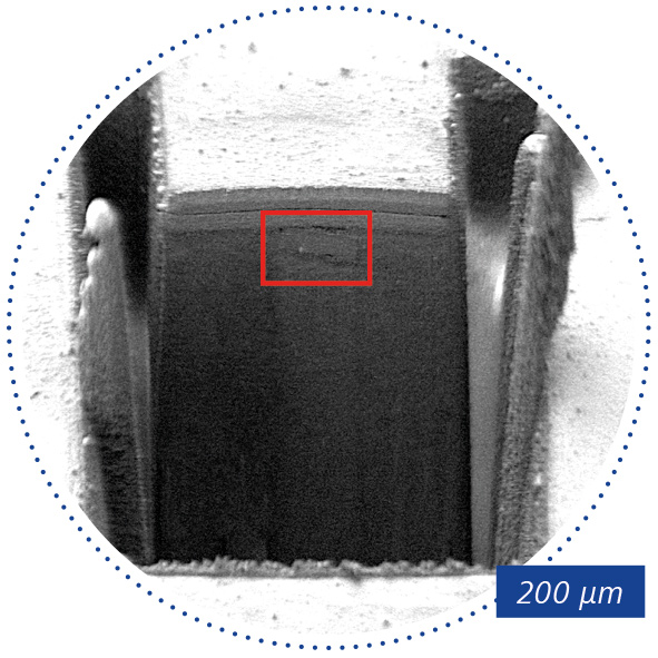


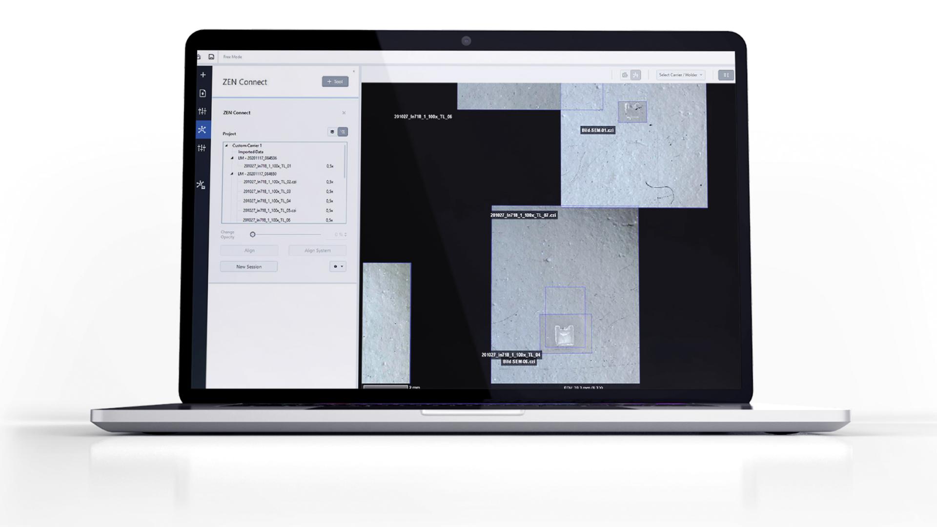

Překrytí laserově frézované spáry na snímku oblasti zájmu ze světelného mikroskopu; SEM, SESI, 450x.